
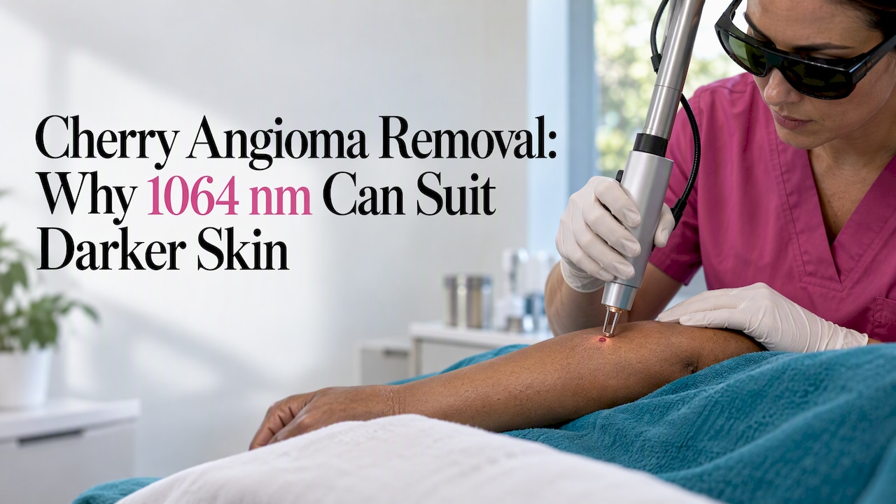

Cherry angiomas are benign clusters of dilated blood vessels, and professional removal can clear treated spots, though the number of visits varies. The main options are pulsed dye laser, long-pulsed 1064 nm Nd:YAG, and electrosurgery, each chosen by lesion size, depth, and skin tone. Lasers tend to heal with lower scarring risk, while electrosurgery is another removal option and, like other methods, can cause scarring.
TL;DR:
- Have a clinician assess spots that grow quickly, bleed without injury, change color, develop irregular borders, or crust before cosmetic treatment begins.
- For deeper skin tones, ask whether 1064 nm Nd:YAG, cooling, conservative settings, and a test spot can lower the risk of lasting pigment changes.
- Cryotherapy is less precise and carries greater risk of lightened skin, while shave excision may suit larger or deeper spots needing one session.
- Keep treated skin clean, avoid picking scabs, protect it from sun, and contact the provider about persistent bleeding, spreading redness, or pus.
Table of Contents
- What a cherry angioma actually is and when removal makes sense
- Professional removal methods compared
- What happens at your appointment and during recovery
- How to choose a provider for cherry angioma removal
- Removal considerations for darker skin tones
- Why diagnosis always comes before treatment
- Getting your cherry angiomas evaluated and treated
- FAQ
- Sources
What a cherry angioma actually is and when removal makes sense
A cherry angioma is a benign cluster of dilated capillaries that shows up as a small, bright red or purple bump, most often on the trunk, arms, or legs. They become more common after age 30, and most people simply accumulate a few over time without any underlying problem, according to the Cleveland Clinic.
Because they are harmless, removing one is almost always an elective, cosmetic decision rather than a medical necessity, the Cleveland Clinic notes. That said, certain changes in a spot deserve a clinical look before anyone reaches for a laser:
- Rapid growth over weeks rather than years
- Spontaneous bleeding without an injury
- Mixed colors, irregular borders, or a crusted surface
Clinicians treat these as signals worth evaluating first, since a handful of other vascular or pigmented lesions can mimic a cherry angioma early on. If a spot behaves normally and simply bothers you cosmetically, removal is a straightforward, low-risk procedure.
Professional removal methods compared
No single method wins across every case. A 2020 systematic review found multiple effective therapies for cherry angiomas, with the right choice depending on lesion size, how raised it is, and the patient’s skin tone.
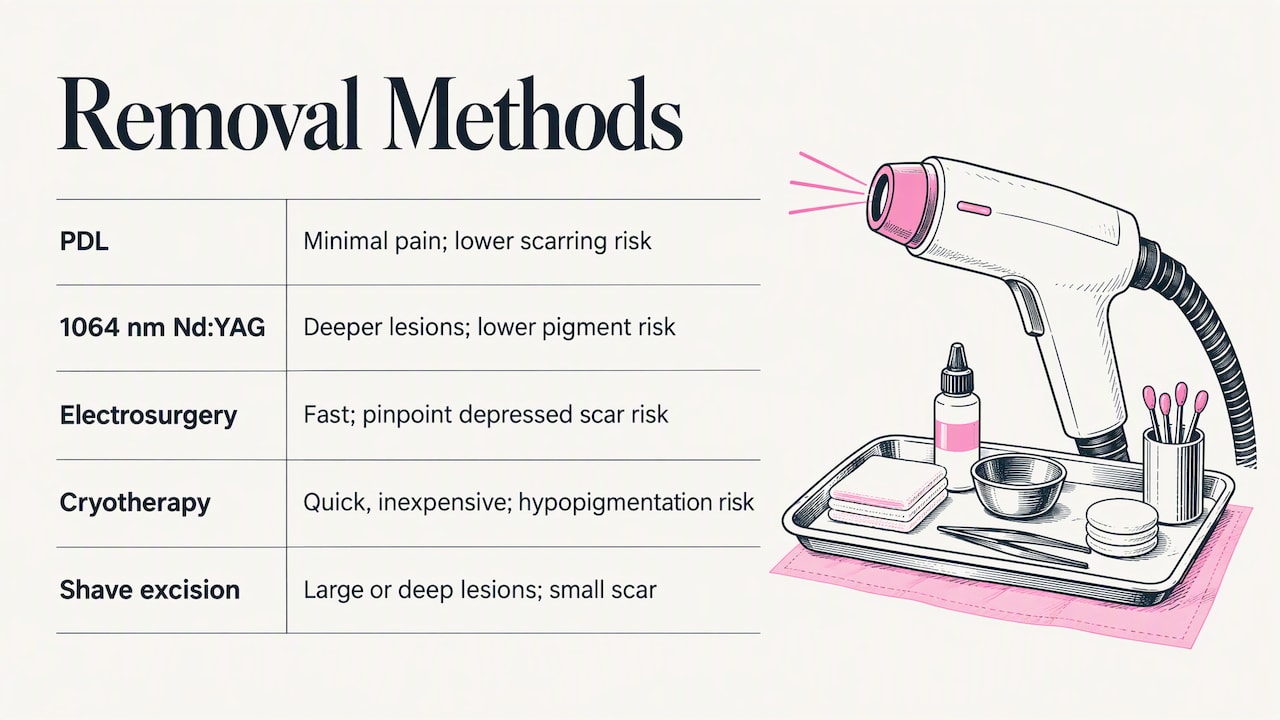
Pulsed dye laser (PDL) targets oxyhemoglobin in the vessel, which absorbs the light and heats the lesion closed without much damage to surrounding skin. PDL is often preferred for its balance of efficacy, minimal pain, and lower scarring risk, per the same systematic review.
Long-pulsed 1064 nm Nd:YAG penetrates deeper than PDL, which makes it a better fit for thicker, more raised lesions. Because the 1064 nm wavelength is less readily absorbed by epidermal melanin, it also carries a lower risk of post-inflammatory pigment changes in darker skin tones compared to shorter wavelengths.
KTP (532 nm) and other short-wavelength lasers are treatment options, though the choice of method depends on skin tone and the risk of pigment changes.
Electrosurgery (electrodesiccation) burns the vessel closed with a fine electrode, which can treat a lesion. It’s fast and effective, though the systematic review notes a somewhat higher risk of small, pinpoint depressed scars compared to laser treatment.
Cryotherapy freezes the lesion with liquid nitrogen. It’s quick and inexpensive but less precise than a laser, and it carries a meaningfully higher risk of hypopigmentation, so it calls for extra caution on skin of color.
Shave excision is reserved for larger or deeper angiomas that don’t respond well to light-based treatment. It trades a small surgical scar for a faster single-session result, and a biopsy may accompany it if there’s any diagnostic doubt about the lesion, per DermNetNZ and a clinical overview of cherry hemangiomas.
A review in PubMed reinforces that no modality is proven definitively superior; the right call depends on matching the device to the spot in front of you.
Pro Tip: Ask which device your provider plans to use and why, since the best choice changes with lesion size, elevation, and your own skin tone.
What happens at your appointment and during recovery
Here’s the general arc of a cherry angioma removal visit, from diagnosis through healed skin:
- Your provider examines the lesion under good light, sometimes with a dermatoscope, to confirm the diagnosis before choosing a method.
- If a spot looks atypical, bleeds without cause, or has grown quickly, a biopsy is prioritized before any cosmetic treatment, following diagnostic triage guidance.
- Treatment itself is generally brief, using topical or local anesthesia as needed.
- With laser treatment, you may see immediate darkening or light bruising at the site as the vessel coagulates; electrosurgery or shave excision closes the area directly.
- Healing varies by treatment method and lesion, and electrocautery or shave sites can take longer to close, per DermNetNZ and the cherry hemangioma overview.
- Full cosmetic settling, where redness fades and texture normalizes, generally takes a few weeks.
Aftercare is simple: keep the area clean, resist picking at any scab, and use sun protection on the healing spot to reduce the chance of lingering pigment change. Call your provider if you notice persistent bleeding, spreading redness, pus, or any unexpected texture or color shift during healing.
How to choose a provider for cherry angioma removal
The right provider examines the lesion before recommending a device, rather than defaulting to whatever equipment happens to be on hand. A clinician who can perform or refer you for a biopsy when something looks off, per DermNetNZ, is taking diagnosis seriously rather than rushing to cosmetic treatment.
A few questions worth asking at your consultation:
- Which devices do you use for cherry angiomas: PDL, Nd:YAG, KTP, or radiofrequency?
- How many sessions do you typically expect for a spot like mine?
- How do you adjust settings or wavelength choice for darker skin tones?
- Can I see before-and-after photos of similar lesions you’ve treated?
- What complication rates have you seen, and how do you manage them?
Beyond the clinical questions, check for clean sterile technique, available local anesthesia for sensitive spots, clear written aftercare instructions, and a defined follow-up policy. If you have several spots to treat, ask whether the practice offers bundled pricing or financing, since multi-lesion removal adds up.
Removal considerations for darker skin tones
Device choice matters more on deeper skin tones, where the wrong wavelength raises the risk of lasting pigment change. Short-wavelength options like 532 nm KTP can carry a risk of pigment changes, particularly on darker skin.
The 1064 nm Nd:YAG laser may be considered for darker skin, as it has shown fewer pigmentary complications.
A few practical adjustments reduce risk further on darker skin.
- Contact or dynamic cooling during the laser pass protects the epidermis from unnecessary heat.
- Conservative fluence settings and longer pulse durations lower the chance of epidermal injury.
- A test spot before treating multiple lesions helps confirm how your skin responds.
Pro Tip: If you have deeper skin tone, ask your provider directly whether they default to Nd:YAG for your lesion type, and follow strict sun avoidance during healing to protect your results.
Why diagnosis always comes before treatment
We’ve seen enough skin over the years to know that a quick glance isn’t a diagnosis. Every spot gets examined under good light before we recommend a removal method, because a lesion that looks like a routine cherry angioma occasionally turns out to be something that needs a closer look first.
We’re also upfront that removing existing spots doesn’t stop new ones from forming elsewhere. That’s part of what we walk through during a consultation, so expectations match reality before anyone books a treatment.
— Chris
Getting your cherry angiomas evaluated and treated
We offer a free consultation where we discuss your skin concerns and recommend a personalized treatment plan. Every plan is personalized rather than one-size-fits-all, and we talk through financing options for anyone treating multiple spots at once.
A few things worth knowing before you book:
- Our skin treatments page outlines our skin treatment options.
- Current prices are on our price booklet, and our membership options are available for clients planning repeat visits.
Ready to find out which method fits your skin? Visit our services page to book your consultation.
This article is general information, not a substitute for advice from a qualified doctor. Consult a qualified healthcare professional about your own circumstances before acting on anything here.
FAQ
Can I remove a cherry angioma myself?
No. Home remedies like apple cider vinegar or tea tree oil don’t actually remove the lesion and risk chemical burns, irritation, infection, or scarring, per the Cleveland Clinic. Professional removal with a laser or electrosurgery is the safe, effective route.
How much does it cost to remove one cherry angioma?
Pricing depends on the device used, lesion size, and number of sessions needed, so it’s best confirmed directly with a provider rather than assumed from a flat rate. Current prices are on our price booklet.
Should you remove cherry angiomas?
Removal is elective unless a spot bleeds, grows rapidly, or raises diagnostic concern, according to the Cleveland Clinic. Many people choose removal purely for cosmetic reasons, and that’s a reasonable call once a provider has confirmed the diagnosis.
Do cherry angiomas mean high estrogen?
There’s no established link confirmed in clinical sources between cherry angiomas and estrogen levels; they are simply benign vascular growths that become more common with age. If you’re concerned about a hormonal connection, that’s worth raising directly with a healthcare provider rather than assuming based on the spots alone.
Sources
- Cherry Angioma: What It Is, Causes & Removal
- Pubmed
- Treatment Modalities for Cherry Angiomas: A Systematic Review
- Dermnetnz


