
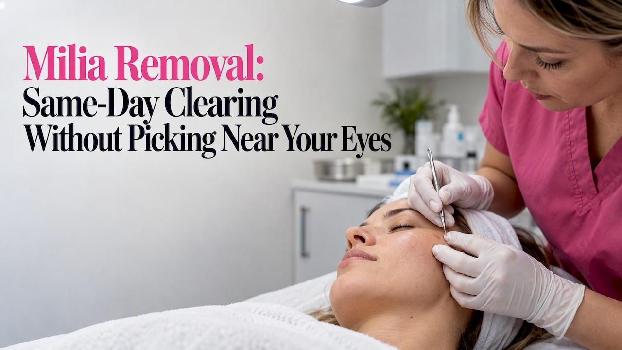

Professional deroofing, a quick in-office procedure where a clinician opens the lesion with a sterile needle and expresses the keratin plug, clears established milia immediately and with the lowest risk of scarring. Topical retinoids and gentle chemical exfoliation help prevent new milia but rarely dissolve bumps that are already there. Avoid picking, poking, or pin-popping milia at home, especially near the eyes, and see a clinician for any eyelid lesion.
TL;DR:
- Neonatal milia usually clear within weeks to months without treatment, while adult milia can persist indefinitely and often require an office procedure.
- Clinicians choose curettage, freezing, or laser based on lesion number, location, and skin type; freezing can leave lighter or darker patches.
- Seek professional assessment if a bump grows quickly, changes color or texture, or several appear within a short period, since other lesions can look similar.
- After treatment, cleanse gently, avoid scrubbing for several days, and use daily sunscreen; treated skin usually looks mostly normal within one to two weeks.
Table of Contents
- What milia are and the different types
- Causes and contributing factors
- Professional removal methods: deroofing, curettage, cryotherapy, and laser
- Preparing for your appointment and caring for your skin afterward
- At-home prevention: retinoids, exfoliation, and routine changes
- When to see a clinician: eyelid caution and red flags
- Possible complications from removal procedures
- Our approach to safe milia care
- Book a consultation for milia removal
- FAQ
- Sources
What milia are and the different types
Milia are small, benign cysts that form just under the top layer of skin when keratin, a protein your skin naturally produces, gets trapped instead of shedding normally. They show up as firm, pearly white or yellowish bumps, usually small and less than a few millimeters wide, most often across the cheeks, nose, and around the eyes. Unlike acne, they have no visible pore or opening, which is part of why squeezing does not work.
Clinicians group them by cause and presentation. Primary (adult) milia appear on otherwise healthy skin with no clear trigger. Neonatal milia show up on newborns and typically clear on their own. Secondary milia follow skin trauma, burns, blistering, or heavy topical products. Milia en plaque is a rarer, clustered form that can appear on the eyelids or behind the ears. Because other bumps, including whiteheads and sebaceous cysts, can look similar, a clinician’s exam helps confirm what you are actually dealing with before you try any treatment.
Causes and contributing factors
Milia form when keratin builds up beneath the skin’s surface instead of shedding the way it normally would, creating a small trapped pocket rather than an open pore. According to StatPearls, these are benign subepidermal keratin-filled cysts that are common and often self-limited.
Several things make that buildup more likely. Heavy, occlusive creams and balms, topical steroids, sun damage, and skin trauma from burns, friction, or cosmetic procedures can all interfere with normal shedding. Skin turnover also slows with age, which is part of why adult milia tend to be more stubborn than the version seen in infants.
That difference matters for how you handle them. Congenital and neonatal milia typically resolve within weeks to months without any treatment, per StatPearls. Acquired adult milia behave differently: they can persist indefinitely and often need an in-office procedure to actually clear rather than fading on their own.
Professional removal methods: deroofing, curettage, cryotherapy, and laser
A handful of clinical techniques account for how dermatologists and medical aestheticians actually clear milia, and the right one depends on lesion number, location, and your skin type.
- Deroofing or manual extraction: a clinician uses a sterile needle or blade to make a tiny opening over the cyst, then expresses the keratin plug, clearing the lesion the same day.
- Curettage: a small surgical instrument scrapes out the contents of the cyst, sometimes paired with light electrodessication to seal the area; healing generally involves a small scab over several days.
- Cryotherapy: brief freezing with liquid nitrogen destroys the cyst wall, an option when there are multiple lesions spread across a treatment area.
- Laser or electrosurgery: targeted energy vaporizes or disrupts the cyst, typically reserved for lesions that are stubborn, numerous, or in areas where precision matters.
According to Cleveland Clinic, treatment options include topical retinoids, in-office surgical removal, cryotherapy, chemical peels, and in some cases electrodessication or laser, while retinoids and exfoliants work better for prevention than for clearing lesions that already exist.
Sterile technique matters across every one of these methods. Milia have no pore to open, so an inexperienced attempt, at home or otherwise, tends to either fail outright or cause unnecessary trauma to surrounding skin. An experienced clinician also distinguishes milia from look-alike lesions before touching anything, which lowers the odds of scarring or a missed diagnosis.
Pro Tip: Ask your clinician which method they recommend for your specific lesion count and location before the appointment, so you know what to expect and can plan around any downtime.
Preparing for your appointment and caring for your skin afterward
A little preparation makes the visit faster and the healing smoother.
- Share your history. Tell your clinician about any skin conditions, medications, or recent procedures, and ask whether to pause retinoids or exfoliants beforehand.
- Expect minimal discomfort. Most milia removal needs no anesthesia; for sensitive areas or multiple lesions, a clinician may use a topical numbing cream.
- Plan for a short visit. Deroofing a handful of lesions usually takes only a few minutes per site, with sterile instruments used throughout.
- Cleanse gently afterward. Use a mild, fragrance-free cleanser and avoid scrubbing the treated area for several days.
- Protect the spot from sun. Fresh skin is more prone to discoloration, so daily sunscreen matters while it heals.
- Watch for infection signs. Increasing redness, warmth, swelling, or discharge should be reported to your clinician promptly; otherwise, expect the area to look mostly normal within one to two weeks.
At-home prevention: retinoids, exfoliation, and routine changes
Home care will not remove milia that are already formed, but it meaningfully lowers how often new ones show up.
- Use a gentle, non-stripping cleanser twice daily rather than anything abrasive.
- Add a measured chemical exfoliant, an AHA like glycolic acid or a BHA like salicylic acid, a few nights a week to encourage normal shedding.
- Switch to lighter, non-comedogenic moisturizers instead of heavy balms that can trap keratin near the surface.
- Apply sunscreen daily, since sun damage is one of the known contributors to milia formation.
Topical retinoids, whether over-the-counter adapalene or prescription tretinoin, are one of the more consistently recommended prevention tools because they speed up cell turnover and help the skin shed keratin normally instead of trapping it. Start two or three nights a week and build up gradually to avoid irritation, and introduce any new acid or retinoid one product at a time rather than all at once.
The skin around the eyes is thinner and more reactive, so keep strong acids and retinoids away from that area unless a clinician has specifically guided you otherwise.
Pro Tip: Introduce one new active ingredient at a time and wait a couple of weeks before adding the next, so you can tell what’s actually working and what’s irritating your skin.
Do not poke, scrape, or use pins, tweezers, or extractor tools on milia at home. There is no pore to open, so these attempts almost always fail and often cause more damage than the bump itself.
When to see a clinician: eyelid caution and red flags
Any milia on or near the eyelid should go to a clinician rather than a home remedy. Dermatologists caution against at-home popping specifically because the periorbital area carries a high risk of scarring or infection.
See a professional sooner rather than later if a lesion grows quickly, changes color or texture, multiples appear in a short window, or any bump looks different from a typical milium. A dermatologist handles most cases, while lesions right on the eyelid margin may warrant an ophthalmologist’s input given how delicate that tissue is.
Possible complications from removal procedures
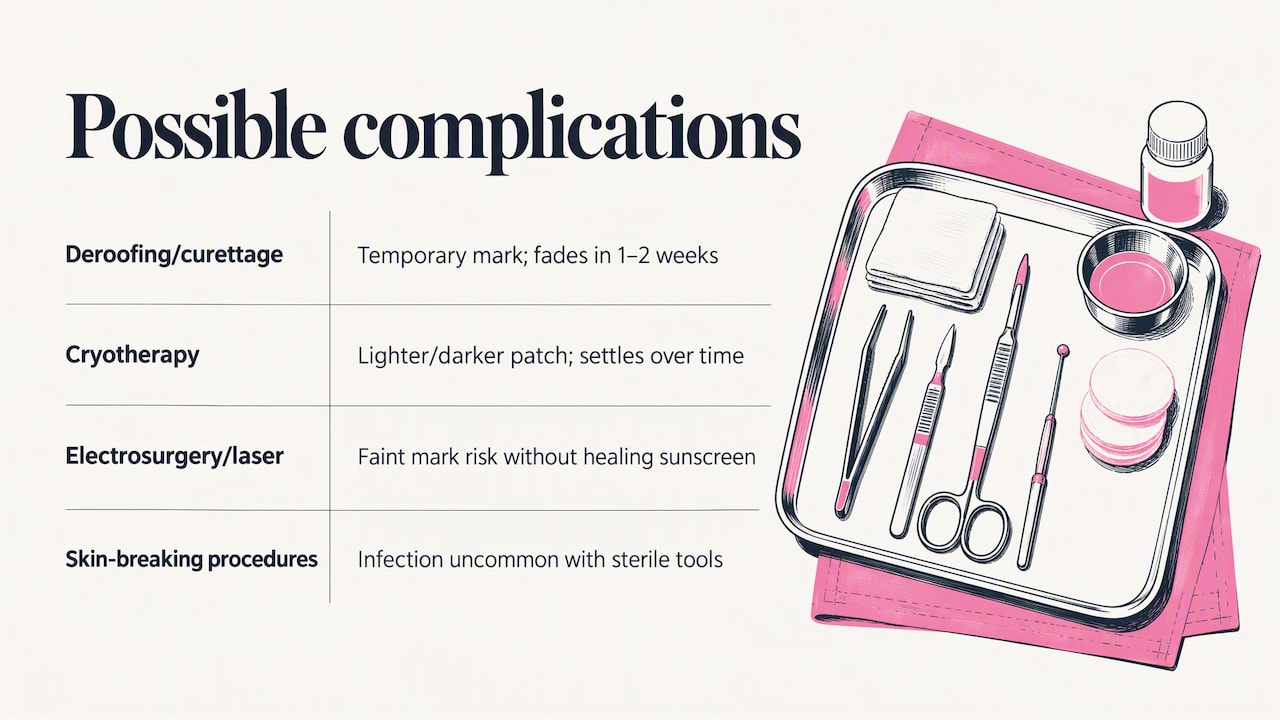
Even straightforward procedures carry a few outcomes worth knowing about ahead of time. Deroofing and curettage can leave a small, temporary scab or pinpoint mark that typically fades within one to two weeks. Cryotherapy occasionally causes a lighter or darker patch of skin at the treatment site, more noticeable in some skin tones than others, which usually settles over time.
Electrosurgery and laser methods carry a modest risk of a faint mark if the area is not protected from the sun during healing, which is why aftercare instructions emphasize sunscreen. Infection is uncommon when sterile instruments are used but remains possible with any skin-breaking procedure, so watch for increasing redness, swelling, or discharge in the days after treatment.
These outcomes are far less likely when the procedure is done by a trained provider with proper sterile technique, which is part of why Cleveland Clinic and other clinical sources consistently steer patients away from DIY extraction and toward in-office care. For anyone unsure whether a bump is milia versus another type of cyst or reaction, a dermatologist backed checklist on distinguishing common skin bumps can help frame the right questions to ask before any treatment.
Our approach to safe milia care
We treat milia removal as a precision procedure, not a quick fix: sterile technique and a consultation before any instrument touches your skin. We build a personalized aftercare plan for every patient, because how you treat the skin afterward matters as much as the extraction itself.
— Chris
Book a consultation for milia removal
If you are dealing with milia that have not budged on their own, we offer in-office evaluation and removal using sterile, clinician-led technique rather than guesswork at home. Our free consultation covers which method fits your skin and lesion pattern, plus a look at the SILVER, GOLD, and PLATINUM membership options if you are interested in ongoing skin care beyond a single visit.

- A consult walks through your history, the lesion itself, and realistic timing for results.
- Membership plans run at various price points per year depending on tier, with financing options potentially available for other treatments.
- Complementary services like facial treatments and Hydrafacial can support overall skin texture alongside milia care.
Ready to get started? Visit our services page to book your consultation.
This article is general information, not a substitute for advice from a qualified doctor. Consult a qualified healthcare professional about your own circumstances before acting on anything here.
FAQ
Can I remove milia myself?
Removing milia at home is not recommended because the cysts have no pore to open, which makes DIY attempts with pins or extractor tools largely ineffective and prone to scarring or infection. A clinician’s sterile deroofing procedure is the standard, effective way to clear established milia.
What is the root cause of milia?
Milia form when keratin becomes trapped beneath the skin instead of shedding normally, often linked to heavy occlusive products, sun damage, skin trauma, or slower cell turnover with age. StatPearls describes them as benign keratin-filled cysts that are common and often self-limited in infants but can persist in adults.
Can milia be permanent?
Adult milia can persist indefinitely without treatment, unlike neonatal milia, which typically resolve within weeks to months on their own, according to StatPearls. Professional removal clears existing lesions immediately, while prevention habits help reduce new ones from forming.
How can I remove milia from my face?
The most reliable option is an in-office deroofing or manual extraction, where a clinician opens the lesion with a sterile instrument and expresses the keratin plug. Cleveland Clinic also lists cryotherapy, chemical peels, and electrodessection or laser as options a provider may choose based on your lesions.


